


The ankle joint is often injured because it bears heavy loads. A doctor can diagnose ankle arthrosis based on symptoms and prescribe treatment. This disease does not depend on age and gender; the tissue becomes thin and destroyed, which can cause deformities.
Arthrosis affects 12% of the population, and more often the disease affects women of retirement age.
As mentioned, the ankle can withstand a large load. It holds the body upright and allows one to move. Its violation changes the normal way of life.
Arthrosis of the ankle joint (symptoms and treatment may vary) is a chronic disease in which irreversible processes occur in the cartilage.
This disease occurs gradually. A healthy person has a smooth articular surface. When overloaded, it promotes easy sliding during physical activity.
With arthrosis, tissue nutrition and metabolism decline. The outer surface of the joint changes, becomes rough, contact cartilage, and inflammation appears. When a person lifts a heavy object, the load falls on the bones, which leads to degenerative disorders.
If treatment is not started, severe pathology develops. In the next stage, the cartilage and tissues are affected, the synovial membrane is irritated, and the joint loses stability. In this case, the support function suffers, movement becomes difficult.
Type
There are several types of arthrosis depending on various criteria:
- cause of the incident (primary, secondary);
- stage of arthrosis;
- localization of pathology;
- forms of localization (general and local);
- disease course (acute and chronic).
| Classification criteria | Types of arthrosis |
|---|---|
| place of manifestation | knee, wrist, ankle, elbow, shoulder and cervical arthrosis. |
| cause of incident |
|
| localization |
|
| disease course |
|
Ankle arthrosis is divided into primary (degenerative process begins in healthy cartilage due to excessive physical activity) and secondary (diagnosed destruction, dystrophic changes appear in the cartilage tissue).
Rank and degree
Arthrosis of the ankle joint (symptoms and treatment directly depends on the patient's age) can occur in many ways. For some people, years pass from the onset of the first symptoms to the critical stage, while for others the disease progresses rapidly.
It depends on the age and concomitant diseases when therapy begins. As ankle arthrosis progresses, symptoms become more pronounced.
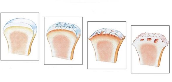
There are 4 stages of the disease.
- The first stage is often overlooked. Main symptoms: stiffness that occurs in the morning, characteristic dryness when walking. Pathogenic changes are not revealed in the image; the destruction process has already begun.
- Stiffness in the morning continues for a longer time. It will take 20-30 minutes to develop the legs. Some patients experience lameness. On the x-ray you can see stage 2 pathology by bone growth and bone displacement.
- In stage 3, symptoms become more pronounced. Painful sensations appear in a calm state, the patient cannot do without painkillers. The lameness becomes noticeable and crutches are sometimes required. Joints swell, change, muscles become thinner and decrease in volume. The joint space narrows, as can be seen on x-rays, and osteophytes form.
- The last stage develops in the absence of treatment. Cartilage is destroyed, the surface of the joint grows together. The patient cannot walk.
There are several stages of arthrosis:
- First degree– X-ray does not show any changes or articulation. There is some morning stiffness. At this stage it is necessary to start treatment.
- In the second degreeactivity becomes difficult, a pulsating sound is heard when walking, swelling is observed. X-rays show a decrease in the interarticular space. The person is weak and the morning cramps are longer.
- In the third stageobvious crusarthrosis, joint deformation. Muscles become more atrophied, movement becomes limited. Constant rest is required. The pain does not go away even in this situation.
- In the last degreeThere is practically no shared space, activities are almost impossible. X-ray allows you to diagnose a large number of osteophytes. Only surgical intervention is prescribed.
Ankle arthrosis appears gradually, so treatment should begin when the first symptoms appear to prevent the condition from worsening and complications from occurring.
symptoms
Arthrosis of the ankle joint is characterized by several symptoms (it affects the treatment method):
- The pain is initially mild and occurs only during physical activity. Over time, the pain becomes stronger and bothers you while resting;
- with injuries and dislocations, swelling and manifestations of inflammation appear, and there is an increase in temperature in the area of the injury;
- click "dry" accompanied by pain;
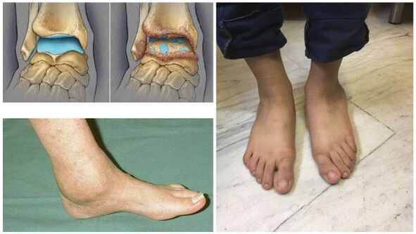
- dislocation, when the cartilage tissue becomes thinner and deteriorates, the joint loses stability. Bones are displaced and fall from the joint capsule;
- joint stiffness;
- when walking, one gets tired quickly;
- in the last stage the joint becomes deformed.
If at least one symptom occurs, you should immediately see a doctor.
Because of appearance
Arthrosis of the ankle joint (symptoms and treatment often due to age-related changes) affects the older generation. Recently, pathology has been observed among young people.
The provoking factors are:
- injuries, dislocations and bruises;
- age-related disorders of the joints and ligaments;
- inflammatory process;
- overweight;
- violation of metabolic processes;
- congenital foot deformities and flat feet that arise during life;
- hereditary predisposition;
- excessive physical activity;
- wearing uncomfortable shoes;
- diseases of the endocrine system;
- osteochondrosis.

Less synovial fluid is produced, resulting in less nutritious cartilage. The joint space narrows, which can cause bone fusion. Crusarthrosis occurs, which cannot be reversed. Even so, treatment should be prescribed immediately to prevent the development of the disease.
Diagnostics
Diagnosis of arthrosis consists of studying existing symptoms and data obtained from research. Since there is no test that can clearly determine the pathology, doctors recognize laboratory methods as not effective enough.
During remission, indicators are normal; during relapse, blood tests show increased ESR and c-reactive protein levels. This means that the pathology has already begun.
To confirm the diagnosis, instrumental methods are used:
- Easyradiographyis the most reliable method. Muscles do not see X-rays equally: soft ones transmit them, while hard ones absorb them. The study revealed the disease itself and its consequences.
The image allows you to analyze the condition of the bone surface in the joint, the shape, size and location of the structures relative to each other, the condition of the tissue and the size of the joint space. Thanks to this data, the degree of pathology can be determined.
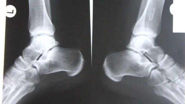
If the ankle is affected, the diagnosis is carried out in lateral, posterior and posterior projections with the foot moved inwards. If there are corresponding symptoms (narrowing of the joint space, osteophytes and other signs), arthrosis is diagnosed.
- Nuclear magnetic resonancedetermine the functional disruption of hydrogen molecules under the influence of a strong magnetic field. Allows you to explore areas of the body that contain water.
The dark colors in the image represent bones, as they contain less water, and muscles, nerves and discs appear lighter. Diagnostics reveal even minor disturbances in bone tissue and joints. This procedure is indicated before joint replacement. The only negative is the high cost of diagnostics.
- Magnetic resonance imagingvery accurately examines the structure of joint ligaments, muscle tissue and cartilage. Thanks to the study, a specialist can assess the condition of the lower leg joints, which makes it possible to identify pathology at an early stage of its development. The procedure is painless and takes about 30 minutes.
During the procedure, strong radio waves and magnetic radiation affect the person. Keep in mind that magnetic fields are dangerous for physiological conditions. MRI is prohibited in case of neuropsychic disorders, pregnancy and the presence of metal objects in the body.
- Ultrasoundenable accurate diagnosis. The device produces waves that are reflected from the tissue and recorded on a screen. The doctor examines the image and makes a diagnosis. For image clarity, a gel is used that expels air and ensures easy movement across the surface.
The advantages of this procedure are health safety, reasonable price and high accuracy.
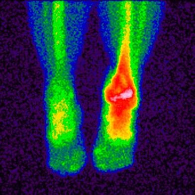
- Bone scintigraphy– studies that make it possible to determine pathological disorders in bones using isotopes. A special material containing labeled atoms is injected into the patient's body. Pathological areas are divided into cold and hot.
In the first there is no isotope, the blood flow to them is weaker, and they are not detected during scanning. This includes places where malignant tumors have appeared. In hot areas, isotopes are collected more actively and are clearly detected during scanning. This area indicates the occurrence of an inflammatory process.
This study makes it possible to separate arthrosis from similar diseases with similar clinical signs; based on the results, the doctor makes a prognosis and prescribes treatment.
The main contraindications for the study are carrying children, breastfeeding and taking drugs containing barium.
- Joint punctureis a procedure in which the doctor inserts a needle into the joint cavity to collect synovial fluid for analysis.
This biomaterial continues to be studied in the future; based on the results, the specialist determines the characteristic features of the disease and the stage of its development. For ankle arthrosis, a puncture is made anteriorly between the outer ankle and the long extensor digitorum tendon.
When you see a doctor
If treatment for arthrosis is not started on time, inability to work and sometimes disability occurs. Some patients do not rush to seek help because they do not know which doctor to make an appointment with. At the first symptoms, you need to visit a rheumatologist who diagnoses dystrophic and inflammatory changes in the joints.
You should contact him if:
- there is discomfort and pain in the joints after excessive load, at the end of the working day;
- it is difficult to find a comfortable position for your legs at night;
- joints swell, skin turns red;
- there is a sharp pain, it is difficult to move;
- pulsating and clicking sounds appear;
- joints become deformed.
With the help of modern diagnostic and therapeutic techniques, it is possible to avoid surgical intervention and preserve joint function.
Prevention
Arthrosis of the ankle joint (symptoms and treatment can be checked with a doctor) can be prevented.
To avoid arthrosis, experts recommend following certain rules:
- wear comfortable shoes that fit properly and without heels;
- maintain proper nutrition, drink enough clean water;
- choose the appropriate vitamin and mineral complex;
- exercise;
- take a walk in the fresh air more often;
- avoid excessive pressure on the feet;
- avoid hypothermia;
- regularly observed by a doctor;
- abandon bad habits;
- do a set of exercises to warm up the ankle joint.

It is very important to adjust your diet. Nutritionists have agreed on a menu that will prevent the aggravation of the disease and saturate the body with the necessary substances.
- You need to eat often and in small portions.
- Drink at least 2 liters of clean water.
- Avoid sweet and salty foods.
- Do not eat food 4 hours before going to bed.
- Steam, bake, boil food.
Fasting and strict diets for arthrosis are strictly prohibited to prevent the leaching of calcium necessary for the restoration of bones and cartilage.
Treatment methods
Once the diagnosis is confirmed, treatment must begin immediately. It is impossible to get rid of arthrosis completely, the main thing is to slow down the destructive process and increase the period of remission. Various techniques are used for this purpose.
Medicines
Various drugs are used to treat arthrosis:
- Anti-inflammatoryand pain relievers remove the source of inflammation and relieve pain. Tablets and ointments are used. The sooner anti-inflammatory drugs are taken, the greater the chance of saving the joint.
- Glucocorticoidsused if the above medicine does not bring the desired result. They are produced in the form of an injectable solution and injected into the joint.
- Chondroprotectorsneeded to slow the process of cartilage destruction.
Treatment regimens and drug dosages are prepared by the doctor based on the severity of symptoms, co-morbidities and other factors. It is strictly forbidden to self-medicate so as not to worsen the situation.
Traditional method
Regarding traditional methods of treating arthrosis, doctors recognize their beneficial properties and positive effects. Traditional medicine is also used as a disease prevention.
The main recipes for the treatment of ankle arthrosis are as follows:
- Burdock leaves are washed clean and rubbed with the soft part on the skin. The plant is fixed with a bandage or cling film and left overnight.
- Heat sea salt (buckwheat, sand) in a pan, pour it into a linen cloth, and apply it to the sore spot. Hold until the salt cools down. This is an effective way to relieve pain.
- Pour triple cologne on the violet, leave it in a dark place for 2 weeks, rub the sore spot twice a day.
- Grind the eggshell into a powder, take 0. 5 teaspoons. before eat.

The use of traditional treatment methods must be agreed with the attending physician. This is not the only measure, but an addition to the main therapy.
Another method
When conservative therapy does not bring positive results, they resort to radical measures - surgery.
As a rule, the indications for surgery are:
- recurrent and primary 3-4 degree arthrosis;
- complications;
- severe and prolonged pain radiating to the knee;
- obvious defects;
- leg muscle paralysis;
- deterioration of joint flexion-extension properties and leg support ability.
For foot arthrosis, the following surgical interventions are used:
- Arthrodesis– surgery to immobilize the joint. Its task is to restore the lost ability to support the limbs. The main disadvantage is the possibility of bones joining together, which leads to immobility, so it is rarely used.
- Arthroscopyis a minimally invasive procedure in which the doctor cuts the joint and inserts an arthroscope. The surgeon conducts a visual inspection and assesses the condition of the intra-articular structures, and, if necessary, removes the damaged part of the joint or a blood clot from the synovial fluid. With this operation the risk of recurrence is too high.
- Endoprostheticscarried out in severe cases. Allows to replace damaged joints in certain parts or completely. Prostheses with modern mechanics are used and last up to 20 years.
The main contraindications for surgery are age under 12 years, fistula in the joint, diabetes mellitus, cardiac dysfunction, and infectious diseases.
Possible complications
If treatment is delayed or absent, the following complications may occur:
- disability;
- irreversible deformation;
- joint inactivity and immobility;
- deterioration of quality and standard of living.
In addition to these complications, the chronic course of the disease is accompanied by pain, discomfort, and the inability to lead an active lifestyle.
To make gymnastics, medicine and folk treatment more effective, it is recommended to use special orthopedic devices that reduce the load on the joints. This includes orthotics and fixation bandages.
The orthosis completely follows the contours of the ankle, increasing range of motion, relieving swelling and pain. A fixation bandage has the same effect as an orthosis. It is made of soft elastic fabric that allows you to fix the joints well. Bandages are used only during the period of remission, when the aggravation passes.
Arthritis of the ankle joint is a serious disease that, if not fully treated, leads to severe consequences and complete immobility of the joint. Diagnosis at an early stage, careful attention to symptoms and competent therapy make it possible to avoid surgical intervention.